

What is a 3D and 4D ultrasound?
An ultrasound tests the high-frequency sound waves, which the human ear cannot hear, by using a transducer to send signals from the abdomen to the computer. Both 3D and 4D ultrasounds are used in obstetric ultrasonography which provides a three dimensional view of the fetus. The difference between 3D and 2D ultrasounds is that in 3D, sound waves are sent to different angles as opposed to 2D, where the sound waves move straight up and down. Both are reflected, and the echoes are then put together by a computer which gives you an image of the fetus and its organs. You are able to see width, height and depth. The advantage of 4D ultrasounds is that they allow this imaging to happen in real time. It just means that are computers becoming smarter and faster.
Ultrasounds can be used to determine the baby’s age, whether or not the pregnancy is high risk, if there are fetal or placental difficulties and if there are problems with the uterus. If an ultrasound is not needed for medical purposes, your insurance company may not pay for the appointment. Contact your insurance provider for information regarding this.
Is it Safe?
Before receiving a 3D or 4D ultrasound, make sure that the facility you use is an accredited ultrasound facility member (AAUIF). This is the first national regulatory association started by facility owners, government officials, doctors and pregnant women who wanted a safe place to receive their ultrasound. Please take note when a facility is not registered. Ultrasounds have no side effects on either the baby or the mother, but it is good to check and see if the facility is following proper protocol.
When Is the Best Time?
Most ultrasounds are performed at 20 weeks to see if the placenta is healthy and the baby is growing properly in the uterus. At this stage in the pregnancy the gender of the baby can be determined, but make sure you let the doctor know whether or not you want to know. They can hide this information from you in the three and four dimensional images as well.
An ultrasound can be performed earlier to see if there is more than one fetus, and to see when the due date will be. Later ultrasounds may be used to make sure the baby and the placenta are healthy, to check the amount of the amniotic fluid and the position and weight of the fetus.
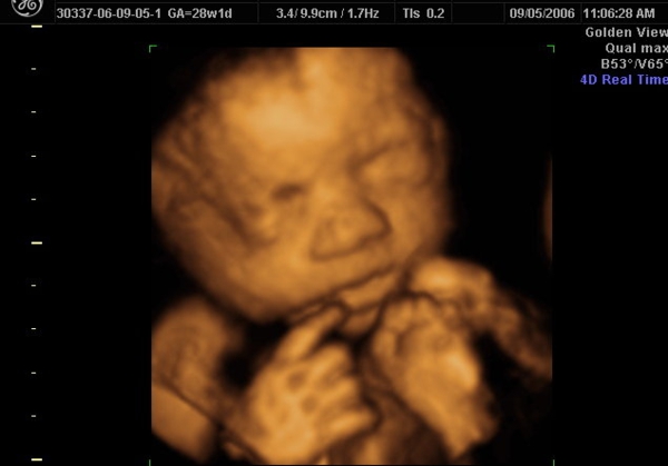
The best time to see facial features in 3D is around 27-32 weeks, but don’t be sad if the baby has their hands in front of their face, this is normal.
Victoria Kunze enjoys writing about 3D ultrasound Los Angeles imaging centers use.